
like humans, dogs may experience a range of eye issues. Among the most frequently encountered eye conditions in dogs are conjunctivitis, cherry eye, cataracts, glaucoma, entropion, dry eye, corneal ulcers, eyelid lumps, pink eye, and keratoconjunctivitis sicca (dry eye).
These ocular conditions can be attributed to various factors, including infections, allergies, insufficient tear production, irritants, scratches, and corneal damage. Some eye issues may also indicate underlying ailments, such as diabetes. If any eye problems are observed in a dog, it is crucial to contact a veterinarian for professional guidance and care.
What is the Collie Eye Anomaly (CEA) in dogs?

Collie Eye Anomaly (CEA) represents a hereditary ocular ailment observed in dogs, impacting both eyes simultaneously. This condition affects very important eye components and these are the:
- Retina – this is situated at the back of the eye, is a layer of cells containing photoreceptors. It converts incoming light into electrical signals sent to the brain for interpretation. Rods and cones are the two distinctive types of light-sensitive cells. Dogs have predominantly rod-dominated retinas, giving them excellent night vision and superior motion detection in low-light conditions compared to humans.
- Choroid – the choroid is a primarily vascular structure that provides nutrients and oxygen to the outer retina. Aside from that, it is critical to ensure that the eye’s temperature and volume are properly regulated. Remarkably, approximately 85% of the blood flow to the eye is directed through the choroidal circulation.
- Sclera – this is a vital component of the eye, and it serves several essential functions in dogs. A delicate membrane covers it called the conjunctiva, extending to the cornea’s edge and lining the interior of the eyelid. Additionally, the sclera provides anchor points for the extraocular muscles responsible for eye movement and contributes to regulating intraocular pressure. Any harm or injury to the sclera can significantly impact a dog’s vision due to its crucial role in the eye’s structure and function.
Origins of CEA can be traced back to a straightforward autosomal recessive genetic anomaly. The onset of this condition stems from irregular eye development, primarily attributed to a deficiency in growth hormone expression within the posterior section of the optic vesicles. This deficiency subsequently influences the differentiation process of other ocular cells.
The prevalence of CEA extends to multiple dog breeds, with a notably higher occurrence among those breeds commonly associated with herding duties.
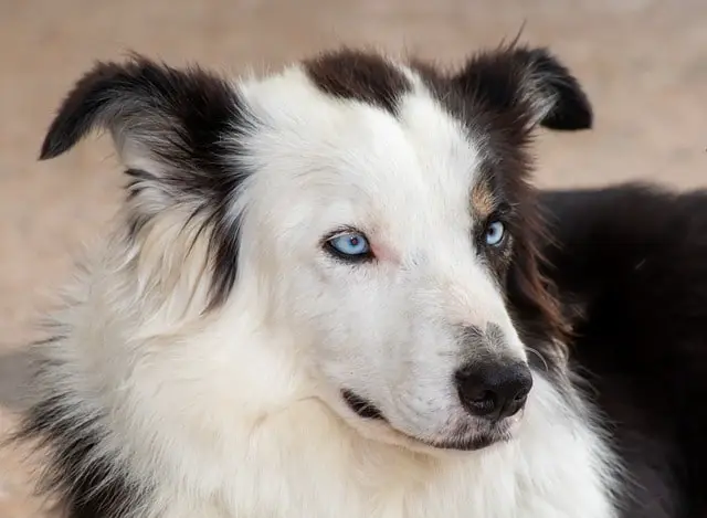
These breeds encompass Collies, Shetland Sheepdogs, Australian Shepherds, Border Collies, and Nova Scotia Duck-Tolling Retrievers.
What are the Clinical Signs of a Collie Eye Anomaly?
In young dogs, clinical manifestations of CEA typically manifest initially. The primary indicator of CEA often observed is impaired vision, with the extent of impairment contingent upon the gravity of the developmental abnormalities. While some dogs maintain normal eyesight, others may experience partial or complete vision loss, particularly if their retinas have become detached.
Additionally, dogs affected by CEA may exhibit other clinical symptoms such as unusually diminutive eyeballs, a sunken appearance of the eye, cloudy eye appearance, and signs of distress or unease when in unfamiliar settings, potentially resulting in accidental collisions with objects and individuals within their home environment.
How is Collie Eye Anomaly (CEA) diagnosed?
Typically, Collie Eye Anomaly (CEA) is identified through a screening process conducted by a veterinary ophthalmologist who examines the front and back of a dog’s eye while the pupils are dilated. There are several methods for diagnosing CEA, which encompass eye examinations, DNA testing, and electroretinograms.
- Eye Examination: This anomaly is often screened for by dog breeders when the canines are between 6 and 8 weeks old. During this examination, a veterinary ophthalmologist administers eye drops to dilate the dog’s pupils and carefully inspects the retina for any discernible alterations indicating the presence of Collie Eye Anomaly. These alterations may include the thinning of tissues surrounding the retina or the occurrence of a coloboma. A coloboma can potentially lead to retinal detachment, ultimately resulting in blindness.
- DNA Test: The sole reliable means of ascertaining whether a Collie possesses a genetically normal eye structure is through a DNA test. It’s important to note that there can be variations even among “Normal” eyes.
- Electroretinogram (ERG): This diagnostic test gauges the retina’s electrical activity when subjected to light stimuli and can identify early indicators of CEA. Typically, this test necessitates sedation or general anesthesia for the dog’s comfort.
Currently, the only effective strategy for reducing the incidence of Collie Eye Anomaly is selective breeding, which considers genetic testing results.
Causes of CEA
A mutation in a specific gene responsible for eye development is the root cause of Canine Eye Anomaly (CEA). This genetic mutation affects the chromosomes governing eye development, leading to inadequate development of the choroid, a network of blood vessels responsible for absorbing scattered light and nourishing the retina.
Furthermore, this genetic alteration can give rise to various eye-related abnormalities, some of which are more severe, such as retinal detachment. Notably, CEA tends to manifest in both eyes, although the severity of its effects may differ.
The prevalence of CEA varies across different dog breeds. In the case of Border Collies, the incidence of this condition is relatively low, ranging from 2% to 3%. Contrastingly, in the United States, it is estimated that up to 95% of Collies either carry the genetic mutation responsible for CEA or have already developed the defect.
Collie Eye Anomaly Types
Various forms of Collie Eye Anomaly can be observed in dogs. CEA is typically categorized as a syndrome due to its encompassing nature and diverse eye irregularities. Predominantly, CEA tends to manifest as choroidal hypoplasia or chorioretinal alterations. Additionally, this condition encompasses defects in the optic nerve or neighboring regions, referred to as coloboma. Further noteworthy variations of CEA involve vascular issues and retinal folds.
Vascular Disease
Vascular disease represents a form of Collie Eye Anomaly, which results in the impairment of the eye’s vascular system responsible for delivering blood to the eye. Consequently, it can give rise to issues related to the eye’s blood circulation, resulting in constriction, inadequate development, or complete absence of these blood vessels.
The presence of vascular disease or tortuous blood vessels can lead to severe complications such as retinal detachment and, in some cases, total vision loss, depending on the extent of the condition’s severity.
Retinal Folds
Retinal folds occur when the developing retina undergoes a folding process, and this anomaly is frequently observed in puppies afflicted with CEA. Nevertheless, in certain instances, retinal folds have the potential to resolve spontaneously.
A veterinary ophthalmologist can identify the presence of retinal folds through an eye examination, during which they will employ an ophthalmoscope to inspect the retina.
Choroidal Hypoplasia
Choroidal hypoplasia represents the most frequently observed anomaly in dogs afflicted with CEA. It refers to thinning the vascular tissue between the retina and the eyeball’s inner wall. A veterinary ophthalmologist can identify this condition during a comprehensive eye examination.
This condition is characterized by a reduction in the growth of blood vessels situated at the rear of the eye, specifically within the choroid. The choroid constitutes a layer of blood vessels positioned between the retina and the inner wall of the eye.
While no curative treatment exists for CEA, surgical intervention may relieve the disease’s symptoms in certain instances.
Coloboma
Coloboma arises due to the incomplete closure of embryonic tissue, leading to the formation of openings or gaps in the optic disc and surrounding regions of the eye. These gaps can develop in either one or both eyes, and their impact on vision varies depending on the extent of the condition.
Collie Eye Anomaly Symptoms
The clinical manifestations of CEA can vary depending on the extent of the developmental defects, but the most prevalent indication is loss of vision. Several observable signs can serve as indicators of CEA and provide a warning to pet owners, including:
- The eyeballs appear to recede within their sockets
- Eyeballs seem smaller than their usual size
- The presence of cloudy eyes
- Impairment of vision
- Complete loss of sight
If you happen to have a Nova Scotia Duck Tolling Retriever, a Shetland Sheepdog, an Australian Shepherd, or a Border Collie, then it is more important for you to ensure that you make yourself aware of these symptoms. Doing so is the only way that you can arm yourself with the right decisions on what move should be done next.
Many responsible dog breeders proactively screen for this abnormality, conducting thorough examinations conducted by a veterinary ophthalmologist when the puppies are between one to two months old. On the other hand, it is unfortunate that, in certain instances, CEA may only be detected once the dog’s vision has been compromised. Having said that, if ever there is any suspicion that your dog is suffering from CEA, it is crucial to promptly seek guidance from a veterinarian to ensure timely diagnosis and treatment.
Smaller Eyeballs
A frequently observed indication of CEA involves the presence of unusually diminutive eyeballs, a condition referred to as microphthalmia. This condition becomes evident to pet owners due to notable eye size alterations. These changes primarily result from choroidal hypoplasia or chorioretinal anomalies, representing CEA’s most prevalent abnormalities. These anomalies adversely affect blood circulation within the choroid, a vascular layer beneath the retina, ultimately reducing eyeball size.
Owners of dog breeds susceptible to CEA should monitor their pets for these telltale signs. If there is any suspicion that their dog may be afflicted with CEA, they must seek advice and consultation from a qualified veterinarian.
Signs of Blindness
The degree of vision impairment experienced by dogs can vary based on the severity of their developmental defects, with some dogs retaining normal vision. Conversely, in more severe instances, CEA can form gaps or depressions in various eye layers, ultimately leading to partial or complete vision loss. Typically, blindness is accompanied by additional indicators such as the presence of cloudy eyes, unusually diminutive eyeballs, and altered behavior that suggests compromised vision.
Dogs affected by CEA may manifest signs of visual impairment, such as inadvertently colliding with objects, hesitance or fear towards venturing outdoors or into unfamiliar environments, and failing to react when fingers are moved close to their eyes.
If a dog owner suspects their pet may be suffering from CEA, it is imperative to seek professional guidance from a veterinarian promptly. A veterinarian can conduct a comprehensive examination of the retina and perform a thorough eye evaluation to determine the extent of the condition and provide appropriate care.
Eyeballs are Sunken
One of the clinical indicators of CEA is the condition known as enophthalmia, characterized by the presence of sunken eyeballs deep within the eye sockets. This occurrence is attributed to choroidal hypoplasia or chorioretinal alterations, frequently observed abnormalities in CEA. These changes impact blood circulation to the vascular layer beneath the retina, referred to as the choroid, resulting in the sunken appearance of the eyeballs.
What are the Treatments for Collie’s Eye Anomaly?
Regrettably, no remedy exists for Collie’s Eye Anomaly. Nonetheless, all hope is not lost, as specific treatments for ocular issues linked to CEA are available. Surgery can be an option to prevent retinal detachment in cases where it poses a concern based on a dog’s specific CEA symptoms.
The surgical approach for reattaching the retina includes two primary methods, with laser surgery and cryosurgery being the most common.
- Laser surgery, recommended by veterinarians in some instances, can address conditions like coloboma, characterized by holes in the lens, choroid, retina, iris, or optic disc.
- On the other hand, cryosurgery is another surgical alternative that employs extreme cold to eliminate unwanted cells or tissues.
It’s essential to understand that the cost of surgery can fluctuate depending on factors such as the severity of the disease and the location of the veterinary clinic. Pet insurance plans might assist in covering some of the expenses associated with CEA treatments. While the precise surgery cost is variable, individuals should anticipate a minimum expense of one thousand dollars per eye.
Nevertheless, surgical intervention for Collie’s Eye Anomaly carries inherent risks. The most severe potential outcome involves recurrent eye bleeding leading to glaucoma, eventual blindness, and the necessity for further surgery to alleviate the dog’s discomfort. This underscores the importance of responsible pet owners conducting thorough research to select a qualified veterinarian for their beloved pets’ surgical needs.
An additional viable treatment option involves conducting annual eye examinations. Dogs with mild cases of CEA may not require any treatment beyond regular eye check-ups to monitor disease progression.
Can the Collie Eye Anomaly (CEA) be Prevented?
A veterinary ophthalmologist can detect Collie Eye Anomaly (CEA) in dogs at approximately two months of age. Preventing CEA involves several steps:
- Firstly, genetic testing allows breeders to identify dogs carrying the CEA gene, helping them avoid pairing such dogs or displaying signs of CEA during eye examinations.
- Secondly, toxin avoidance is crucial in supporting healthy puppy development, involving protecting the mother from toxins, administering proper vaccinations, maintaining a parasite-free environment, and ensuring a nutritious diet.
- Lastly, combining genetic testing with selective breeding is the primary method to reduce CEA transmission. This entails screening dogs for the gene mutation linked to CEA and breeding carriers with tested, clear dogs to prevent the condition while maintaining genetic diversity.
The most effective approach for CEA prevention involves comprehensive eye exams and DNA testing in breeding dogs. Once CEA develops, there is no cure. Pet owners seeking help should:
- Locate a local veterinary ophthalmologist specializing in eye diseases.
- Consult their regular vet for a referral to a trusted ophthalmologist.
- Inquire at nearby animal hospitals or clinics for recommendations.
- Conduct online research or use resources like the American College of Veterinary Ophthalmologists for referrals.
Once a suitable ophthalmologist is found, schedule a comprehensive eye examination for the dog, including retina assessment and potentially a genetic test to confirm CEA diagnosis.